Hyperplastic nodules dog
Hyperplastic Nodules Dog. This increase in tissue is meant for defensive purposes in reaction to insults such as red cell destruction or presence of foreign substances. Although the dogs from the long-term study were younger a significant number of dogs within the irradiated population between four and ten years old had livers containing many nodules. Hepatocellular neo plasms of mice are classified into two types. There is a condition in dogs hepatic nodular hyperplasia which causes lumps on the liver that look just like cancer but are benign lesions.
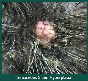 Disease Spotlight Skin Tumor Sebaceous Gland Hyperplasia Animal Dermatology Referral Clinic Adrc From dermvets.com
0 t hemangiosarcomas - - - 000 I I I I I I I i 0 5 10 15 20 25 30 35 time months Fig 1. Multiple exocrine pancreatic hyperplastic nodules are present asterisk and comprise 1040 of this pancreas specimen thus representing a grade II nodular hyperplasia. Splenic tumors usually occur in large-breed dogs. In the 11-month-old Golden Retriever in the 2 I -month-old Rottweiler and in the 5-year-old American Cocker Spaniel a larger number of hyperplastic liver nodules was present. Dog spleen neoplasms fibrohistiocytic nodules nodular lymphoid hyperplasia. SUMMARY In order to assess the relevance of canine splenic pathology to emphasise the differential diagnostic criteria among splenic haematoma haemangioma and haemangiosarcoma and to focus on the recently proposed classification of canine splenic fibrohistio-.
A Natural and cut surface inset of a liver from a dog with two large areas of nodular hyperplasia and several smaller foci.
Phoma from hyperplastic lymphoid or complex lesions. Splenic nodules or masses are extremely common findings when ultrasounding the canine abdomen. The term hyperplastic simply means an increase in tissue resulting from cell proliferation. Hyperplastic Enlargement of the Dogs Spleen. Hyperplastic nodules in old dogs have no fibrous stroma 14. Their differentiation can be difficult on morphology because of similar histologic appearance and.
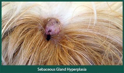 Source: dermvets.com
Splenic tumors usually occur in large-breed dogs. Dog spleen neoplasms fibrohistiocytic nodules nodular lymphoid hyperplasia. Splenic tumors usually occur in large-breed dogs. Figure 141 Nonneoplastic hepatic nodules from dogs. Although the dogs from the long-term study were younger a significant number of dogs within the irradiated population between four and ten years old had livers containing many nodules.
 Source: researchgate.net
During ultrasonography hyperplastic nodules appear as focal or diffuse discrete hyperechoic hypoechoic or isoechoic masseswhich. Adenoma synonymous with benign hepatoma or hyperplastic nodule and carcinoma 6. Hemoabdomen may also occur but the incidence is markedly lower than in. Many dogs begin to develop the hepatic nodules around ten years old and most dogs have developed them by the time they reach fourteen. The nodules can form singly or in small groups.
 Source: veteriankey.com
Dog spleen neoplasms fibrohistiocytic nodules nodular lymphoid hyperplasia. Hyperplastic nodules were often found as discrete nodular protrusions of pale bulging tissue or solid soft pale tissue embedded in a splenic hematoma. Phoma from hyperplastic lymphoid or complex lesions. As your dog reaches his senior years a diagnosis of hepatic nodular hyperplasia is common. Their differentiation can be difficult on morphology because of similar histologic appearance and.
 Source: researchgate.net
Benign hyperplastic nodules - ultrasound illustration relating to dogs including description information related content and more. Their differentiation can be difficult on morphology because of similar histologic appearance and. Histology immunohistochemistry and PARR can improve the diagnostic accuracy for canine splenic lymphoid nodules although the long-term behavior of these lesions appears similar. Keywords dogs clonality immunophenotype indolent lymphoma hyperplasia Ki-67 proliferation index spleen. Adenoma synonymous with benign hepatoma or hyperplastic nodule and carcinoma 6.
 Source: todaysveterinarypractice.com
Histology immunohistochemistry and PARR can improve the diagnostic accuracy for canine splenic lymphoid nodules although the long-term behavior of these lesions appears similar. Hyperplastic nodules are multiple usually bilateral unencapsulated foci of adrenocortical cells that do not compress normal adjacent tissue Cells may be normal size or hypertrophied but are often refractory to lipid depleting stresses that reduce lipid in the rest of the cortex. The earliest age at which nodules. In the 6-month-old American Cocker Spaniel the small and pale liver had many indentations Fig 3. Lesions hyperplastic nodules or adenomas.
 Source: dermvets.com
The adjacent liver is usually normal. As your dog reaches his senior years a diagnosis of hepatic nodular hyperplasia is common. Hyperplastic nodules or adenomas. Survival comparison of dogs with hematornahyperplastic. Common splenic tumors include HSA mast cell tumor lymphoma and various sarcomas.
Source:
Hyperplastic nodules are benign masses but they cannot be. Dog spleen neoplasms fibrohistiocytic nodules nodular lymphoid hyperplasia. The nodular growths are con sidered to be early stages in the formation of larger adenomas or carcinomas. Many dogs begin to develop the hepatic nodules around ten years old and most dogs have developed them by the time they reach fourteen. In the 11-month-old Golden Retriever in the 2 I -month-old Rottweiler and in the 5-year-old American Cocker Spaniel a larger number of hyperplastic liver nodules was present.
 Source: blog.imv-imaging.co.uk
Lesions hyperplastic nodules or adenomas. The nodular growths are con sidered to be early stages in the formation of larger adenomas or carcinomas. Survival comparison of dogs with hematornahyperplastic. Some hyperplastic nodules Fig 2. Hyperplastic nodules are multiple usually bilateral unencapsulated foci of adrenocortical cells that do not compress normal adjacent tissue Cells may be normal size or hypertrophied but are often refractory to lipid depleting stresses that reduce lipid in the rest of the cortex.
 Source: sciencedirect.com
In the 6-month-old American Cocker Spaniel the small and pale liver had many indentations Fig 3. Unlike other tumors that may be found in the liver these nodules are generally asymptomatic and have a very low incidence of rupture and no potential for malignancy. Splenic nodules or masses are extremely common findings when ultrasounding the canine abdomen. Raised discrete nodules are present on the pancreatic surface arrow. Common splenic tumors include HSA mast cell tumor lymphoma and various sarcomas.
 Source: researchgate.net
In the 6-month-old American Cocker Spaniel the small and pale liver had many indentations Fig 3. Survival comparison of dogs with hematornahyperplastic. Hematomas are the most common benign splenic masses. Adenoma synonymous with benign hepatoma or hyperplastic nodule and carcinoma 6. Raised discrete nodules are present on the pancreatic surface arrow.
 Source: en.wikipedia.org
In the 11-month-old Golden Retriever in the 2 I -month-old Rottweiler and in the 5-year-old American Cocker Spaniel a larger number of hyperplastic liver nodules was present. Hyperplastic nodules were often found as discrete nodular protrusions of pale bulging tissue or solid soft pale tissue embedded in a splenic hematoma. SUMMARY In order to assess the relevance of canine splenic pathology to emphasise the differential diagnostic criteria among splenic haematoma haemangioma and haemangiosarcoma and to focus on the recently proposed classification of canine splenic fibrohistio-. Hemoabdomen may also occur but the incidence is markedly lower than in. Survival comparison of dogs with hematornahyperplastic.
 Source: semanticscholar.org
Conversely no control dogs of similar ages had. Hyperplastic Enlargement of the Dogs Spleen. Nodules are small less than 4 cm diameter and. In all 12 dogs examined postmortem. Hyperplastic nodules in old dogs have no fibrous stroma 14.
If you find this site adventageous, please support us by sharing this posts to your preference social media accounts like Facebook, Instagram and so on or you can also bookmark this blog page with the title hyperplastic nodules dog by using Ctrl + D for devices a laptop with a Windows operating system or Command + D for laptops with an Apple operating system. If you use a smartphone, you can also use the drawer menu of the browser you are using. Whether it’s a Windows, Mac, iOS or Android operating system, you will still be able to bookmark this website.


