Mass in stomach dog
Mass In Stomach Dog. May 18 2010 a 15lbs tumor mass was removed from this 13 years old dogs abdomen at the airport animal emergency center in indianapolis on 5172010. Adenocarcinomas for example are among the most common gastrointestinal tumors in dogs and usually develop in the colon rectum and stomach. Most times this tumor is merely a benign fatty mass that can easily be removed allowing your dog a full recovery. Splenic masses are the most common canine abdominal mass seen in veterinary medicine.
 Canine Stomach Cancer Lovetoknow From dogs.lovetoknow.com
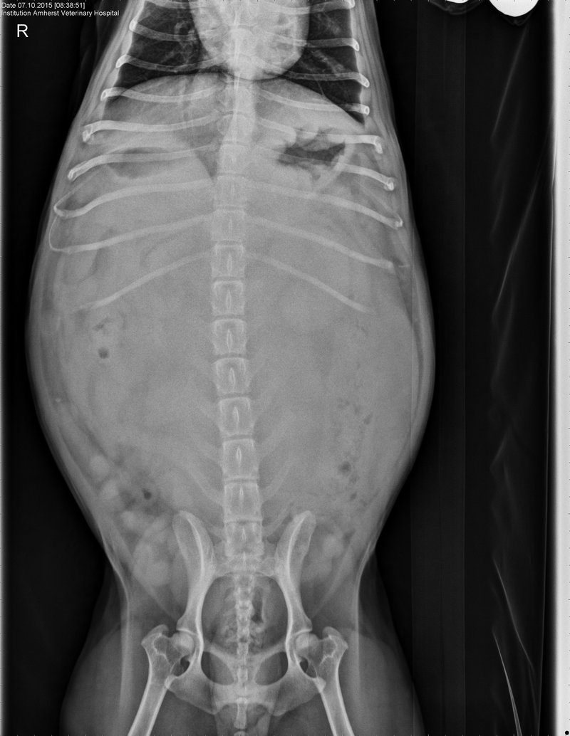
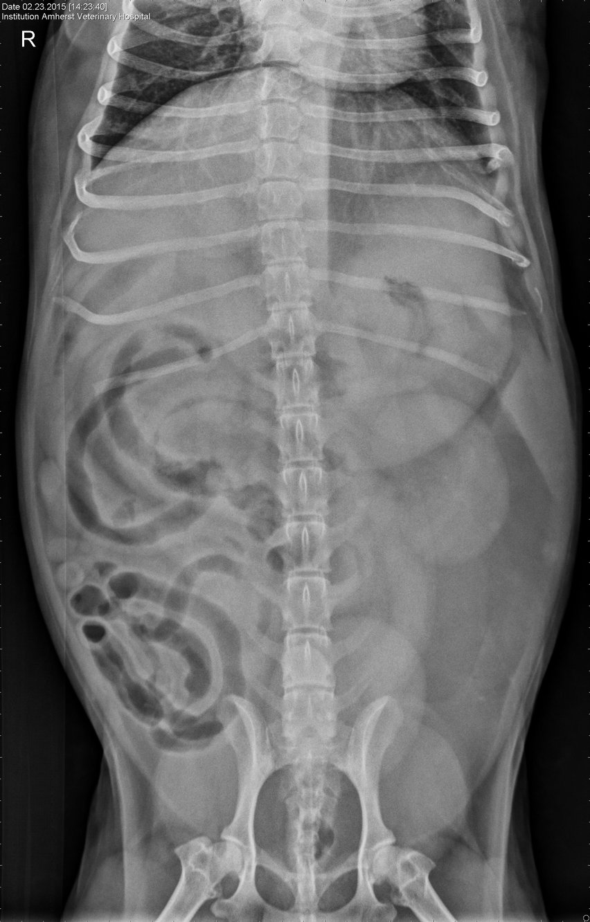
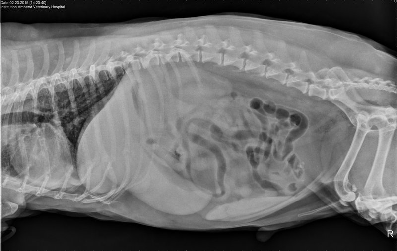
Lumps and bumps can be harmless or can be cause for concern. Intestinal cancer mostly takes the form of a malignant tumor called adenocarcinoma which originates in the epithelial and glandular tissue. Gastrointestinal cancer is fairly uncommon in dogs but it does occur. Benign masses can be hemangiomas hematomas fibromas or leiomyomas which are all fancy medical names for different non cancerous masses that can be found on the spleen. The ventrodorsal abdominal radiograph identifies a round soft tissue opacity in the deep part of the fundus of the stomach concerning for foreign material gastric mass or ingesta. Yes it is an ominous problem but it does not require immediate euthanasia in my view.
Sometimes the stomach and intestines become twisted leading to a life-threatening condition that requires emergency surgery.
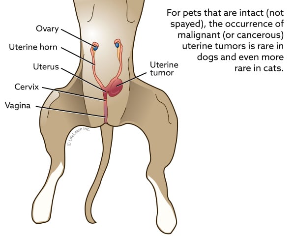
If your vet suspects a mast cell tumor your dog may be treated first with diphenhydramine to minimize the histamine release. Stomach tumors in dogs gastrointestinal tumors in dogs can be of several different types and although the symptoms can often be quite serious not all tumors are. This type of neoplasm can invade a variety of parts of the gastrointestinal system including the rectum. Splenic masses are the most common canine abdominal mass seen in veterinary medicine. Your vet will need to take some tests to accurately diagnose your dogs lump. The most common type of canine cancer affecting the intestinal tract is lymphoma followed by adenocarcinoma.
 Source: amherstvethospital.com
Splenic masses are the most common canine abdominal mass seen in veterinary medicine. Many people complain of stomach ache mostly in the evening or after meals. Focal wall thickening with a loss of wall layering are commonly seen with intestinal focal neoplasia FIGURE 18. The increased risk of Belgian Shepherds for gastric carcinoma and of Siamese cats for intestinal adenocarcinoma and. Whenever the diagnosis of an abdominal mass is made it is important to gain perspective.
 Source: vetfolio.com
As is often the case with malignant splenic masses this dog behaved perfectly normally in the morning but died by mid-day soon after his spleen ruptured. This tumor most commonly occurs in the skin as a rai. Stomach wall thickness in dogs is 3 to 5 mm depending on the location and size of the dog with larger dogs having a thicker stomach wall. 3756 In cats the rugal-fold thickness has been reported as 44 mm 57 26 mm 57 or 20 mm 58 for the interrugal area and 21 mm for the pylorus. The most common type of canine cancer affecting the intestinal tract is lymphoma followed by adenocarcinoma.
 Source: vcahospitals.com
3B and the histopathological results for these specimens indicated hypertrophic gastritis Fig. The spleens function is a red blood cell processing plant. Tumors in dogs abdomen search now. Gastrointestinal GI neoplasms are uncommon in dogs and cats with gastric tumors representing 1 and intestinal tumors 10 of overall neoplasms in the dog and catSpecific etiologic agents for GI neoplasia have not been identified. A gastrointestinal endoscopic examination was performed and a proliferative mass was found in the pyloric outflow tract Fig.
 Source: medvetforpets.com
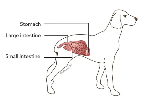
The dogs spleen is a highly vascular organ that sits behind the stomach. Gastrointestinal cancer is fairly uncommon in dogs but it does occur. If your vet suspects a mast cell tumor your dog may be treated first with diphenhydramine to minimize the histamine release. Bernards and German Shepherds large amounts of gas can get trapped in the stomach and intestines and cause serious abdominal distention. Sometimes the stomach and intestines become twisted leading to a life-threatening condition that requires emergency surgery.
 Source: vcahospitals.com
Gastrointestinal tumors can occur anywhere in the gastrointestinal tract but stomach tumors in dogs tend to be more common. Dogs over the age of 9 are most likely to develop the disease with males affected more than females. However the location also depends on the type of tumor. These masses may release histamine when disturbed which can have a negative effect on your dogs body. The dogs name is.
 Source: amherstvethospital.com
Leiomyoma of the Stomach Small and Large Intestine in Dogs. Acid reflux diet myths and natural food solutions. Skin lumps and bumps in dogs what you should know. However the location also depends on the type of tumor. They may recommend a fine needle aspirate and cytology one of the least invasive procedures to evaluate a lump or bump during which a vet uses a small.
 Source: dogs.lovetoknow.com
In most cases the dog will respond within 72 hours and gradually regain strength. The increased risk of Belgian Shepherds for gastric carcinoma and of Siamese cats for intestinal adenocarcinoma and. Black arrows define margins of suspected foreign material. A leiomyoma is a relatively harmless and nonspreading tumor that arises from the smooth muscle of. It commonly affects dogs that are older than six years of age.
 Source: researchgate.net
Gastric lymphoma in dogs and cats can be localized growing as a mass in one location or diffuse spread out as a general thickening of the stomach wall. The stomach and proximal duodenum was markedly distended in the dog with the duodenal mass and regional dilatation of bowel segments possibly indicating delayed passage of. Black arrows define margins of suspected foreign material. Adenocarcinomas for example are among the most common gastrointestinal tumors in dogs and usually develop in the colon rectum and stomach. 14354546 The most common intestinal tumors of dogs are leiomyosarcoma lymphoma and adenocarcinoma.
 Source: vetfolio.com
Stomach wall thickness in dogs is 3 to 5 mm depending on the location and size of the dog with larger dogs having a thicker stomach wall. Treat the abdomen as a cracked egg ready to break at any time. Most times this tumor is merely a benign fatty mass that can easily be removed allowing your dog a full recovery. Tumors in dogs abdomen. This tumor most commonly occurs in the skin as a rai.
 Source: medvetforpets.com
It commonly affects dogs that are older than six years of age. If your vet suspects a mast cell tumor your dog may be treated first with diphenhydramine to minimize the histamine release. They may recommend a fine needle aspirate and cytology one of the least invasive procedures to evaluate a lump or bump during which a vet uses a small. Gastrointestinal GI neoplasms are uncommon in dogs and cats with gastric tumors representing 1 and intestinal tumors 10 of overall neoplasms in the dog and catSpecific etiologic agents for GI neoplasia have not been identified. As is often the case with malignant splenic masses this dog behaved perfectly normally in the morning but died by mid-day soon after his spleen ruptured.
 Source: amherstvethospital.com
Tumors in dogs abdomen. Yes it is an ominous problem but it does not require immediate euthanasia in my view. Leiomyoma of the Stomach Small and Large Intestine in Dogs. Stomach tumors in dogs gastrointestinal tumors in dogs can be of several different types and although the symptoms can often be quite serious not all tumors are. 58 Five wall layers corresponding to the mucosal surface mucosa submucosa.
 Source: petcancercenter.org
Gastric lymphoma in dogs and cats can be localized growing as a mass in one location or diffuse spread out as a general thickening of the stomach wall. Gastric lymphoma in dogs and cats can be localized growing as a mass in one location or diffuse spread out as a general thickening of the stomach wall. They may recommend a fine needle aspirate and cytology one of the least invasive procedures to evaluate a lump or bump during which a vet uses a small. 3AMultiple biopsy samples were taken with grasping forceps Fig. The ventrodorsal abdominal radiograph identifies a round soft tissue opacity in the deep part of the fundus of the stomach concerning for foreign material gastric mass or ingesta.
If you find this site adventageous, please support us by sharing this posts to your favorite social media accounts like Facebook, Instagram and so on or you can also save this blog page with the title mass in stomach dog by using Ctrl + D for devices a laptop with a Windows operating system or Command + D for laptops with an Apple operating system. If you use a smartphone, you can also use the drawer menu of the browser you are using. Whether it’s a Windows, Mac, iOS or Android operating system, you will still be able to bookmark this website.


