Mass on xray of dog
Mass On Xray Of Dog. Depending on the grade of the tumor dogs may live and survive upwards of 22 months or only survive an additional six months. Acute nontraumatic hemoabdomen in the dog. Dyspnea is the most common presenting clinical sign. J Am Vet Med Assoc 20112391325-7.
 Xray In Dogs Vets On The Balkans An Online Journal For Veterinarians From The Balkans From balkanvets.com
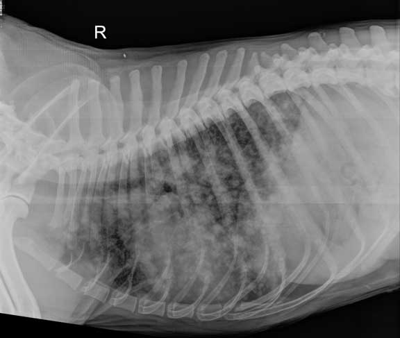
The mass was visible in the same spot in all four xrays confirming its place within the chest in a certain part of the lungs - the right caudal lung lobe. Clinically there was upper respiratory noise and a 32 x 28 x 27 cm ovoid mass involving the epiglottis was observed. This test can also be helpful in cases of unexplained fever. Thymoma in an 11-year-old dog. Once ready the x-ray will be triggered where it will take images of the area in a variety of grey shades but dense tissue will come up white. Before knowing if the procedure would help the type of tumor your dog has would have to be determined.
This test can also be helpful in cases of unexplained fever.
A retrospective analysis of 39 cases 1987-2001. Bloodwork would be helpful because it can tell you about possible infections that could cause a mass and also will tell you something about her liver. When a dog needs an x-ray he is placed on a table and positioned in such a way that the area that the veterinarian is interested in seeing will be targeted by the x-ray beam. Thymoma in an 11-year-old dog. Clinically there was upper respiratory noise and a 32 x 28 x 27 cm ovoid mass involving the epiglottis was observed. The x-ray tube head should be rotated approximately 5 to 10 degrees caudally angle light from caudal to cranial as seen from the dog to prevent superimposition of the femur and tibia at the level of the stifle joint.
 Source: lbah.com
Clinical signs are variable and are related to a space-occupying cranial mediastinal mass andor manifestations of the paraneo-plastic syndrome. Your dog may need to be re-positioned to allow all the necessary angles to be covered. Other sites of metastasis include lymph nodes bone or other organs all of which can be evaluated by radiographs. Clinical signs are variable and are related to a space-occupying cranial mediastinal mass andor manifestations of the paraneo-plastic syndrome. An x-ray can differentiate BONE as white but other soft tissue organs including fluid such as urine in the bladder as gray.
 Source: petcancercenter.org
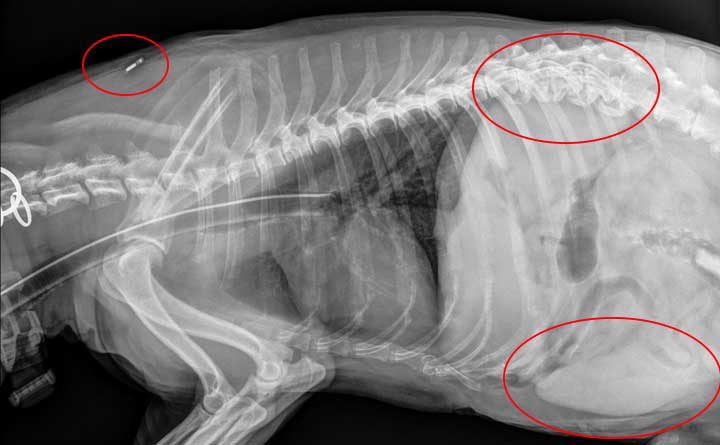
A lateral X-ray of a dogs chest and cranial abdomen. ABSTRACT Primary chondrosarcoma was found in the epiglottis of a 6-year-old neutered male Boxer Cross. Bloodwork would be helpful because it can tell you about possible infections that could cause a mass and also will tell you something about her liver. In your 14 year old dog you mention that it is a gray mass along the right side of the body and appears as a gray shadow over the internal organs. If a pet comes in limping X-rays of the affected limb s are taken to look for broken bones arthritis hairline fractures or other causes.
 Source: researchgate.net
The best test to know exactly what this mass involves is an ultrasound. Adrien Hespel DVM MS DACVR. 50 of dogs with this type of cancer live at least one year beyond the removal of the mass. The normal thymus is visualized in the cranioventral mediastinum in young dogs as an inverted wedge shape known as a sail sign Figure 1. An x-ray cannot tell you the color of the internal organs.
 Source: shutterstock.com
Dogs that present with primary lung cancer with just a single small mass in their lungs that has stayed contained are good. The tube angle is dependent on the muscle mass of the dog when the limb is in an extended position. This will include a chemical blood profile a complete blood count a urinalysis and an electrolyte panel. Abdominal X-rays are indicated to evaluate dogs with abdominal symptoms such as vomiting retching constipation or diarrhea. Dogs that have this procedure survive on average for nearly two more years.
 Source: medvetforpets.com
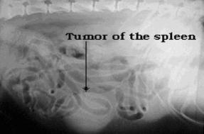
Adrien Hespel DVM MS DACVR. If her liver values are high then this mass may be affecting her liver and that could be a very serious sign. A chest x-ray and ultrasound imaging will allow your veterinarian to visually examine the heart so that a complete assessment can be made of the heart and any masses that are present within it. Atlas of anatomy on x-ray images of the dog. In VD abdominal radiographs of the dog it is the proximal extremity of the spleen that typically is seen Figure 7-13.
 Source: lbah.com
Acute nontraumatic hemoabdomen in the dog. The head is towards the left. Other imaging modalities would be needed to interpret this finding correctly because a mass effect between the gastric fundus and kidneys in lateral radiographs could represent an abnormal mass. Other sites of metastasis include lymph nodes bone or other organs all of which can be evaluated by radiographs. Again this is due to the portion of the proximal.
 Source: drphilzeltzman.com
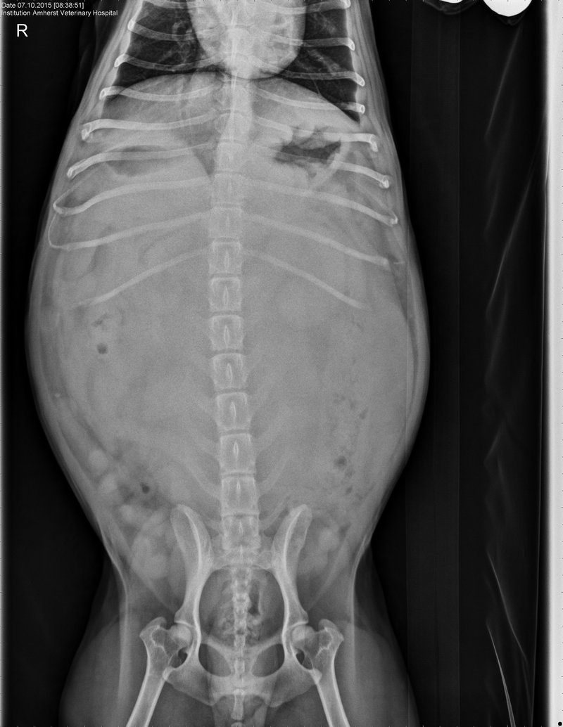
The x-ray tube head should be rotated approximately 5 to 10 degrees caudally angle light from caudal to cranial as seen from the dog to prevent superimposition of the femur and tibia at the level of the stifle joint. The best test to know exactly what this mass involves is an ultrasound. Ventrodorsal abdominal radiograph of a dog with two-day history of vomiting. The tube angle is dependent on the muscle mass of the dog when the limb is in an extended position. No abnormalities were detected on radiographic examinations X-ray of.
 Source: medvetforpets.com
Again this is due to the portion of the proximal. Other sites of metastasis include lymph nodes bone or other organs all of which can be evaluated by radiographs. The head is towards the left. A lateral X-ray of a dogs chest and cranial abdomen. As a rule radiation is administered daily for 5 days per week 2 rest days until 18 to 21 treatments have been completed.
 Source: amherstvethospital.com
Mass-to-splenic volume ratio and splenic weight as a percentage of body weight in dogs with malignant and benign splenic masses. 50 of dogs with this type of cancer live at least one year beyond the removal of the mass. This is a radiograph of the abdomen of a normal cat that is laying on its right side. In VD abdominal radiographs of the dog it is the proximal extremity of the spleen that typically is seen Figure 7-13. Thoracic radiographs of 75 dogs and cats with mediastinal andor pulmonary masses identified on CT were reviewed.
 Source: balkanvets.com
An x-ray cannot tell you the color of the internal organs. Therefore radiographs of the chest commonly referred to as thoracic radiographs are often performed in dogs with a confirmed or suspected diagnosis of cancer. Dogs that present with primary lung cancer with just a single small mass in their lungs that has stayed contained are good. A retrospective analysis of 39 cases 1987-2001. Radiographic studies were anonymized randomized and reviewed twice by three reviewers.
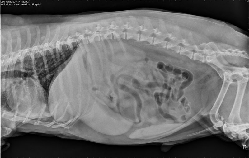 Source: amherstvethospital.com
The x-ray tube head should be rotated approximately 5 to 10 degrees caudally angle light from caudal to cranial as seen from the dog to prevent superimposition of the femur and tibia at the level of the stifle joint. In VD abdominal radiographs of the dog it is the proximal extremity of the spleen that typically is seen Figure 7-13. An x-ray cannot tell you the color of the internal organs. Given lulus age and how the mass looks on the xray - it is very much likely that this a tumor. A paraneoplastic syndrome of myasthenia gravis nonthymic malignant tumors andor polymyositis occurs in a significant number of dogs with thymoma.
 Source: greatpetcare.com
Radiographic studies were anonymized randomized and reviewed twice by three reviewers. Again this is due to the portion of the proximal. In general tumors located on the front part of the jaw have a better prognosis. Adrien Hespel DVM MS DACVR. Given lulus age and how the mass looks on the xray - it is very much likely that this a tumor.
If you find this site serviceableness, please support us by sharing this posts to your favorite social media accounts like Facebook, Instagram and so on or you can also save this blog page with the title mass on xray of dog by using Ctrl + D for devices a laptop with a Windows operating system or Command + D for laptops with an Apple operating system. If you use a smartphone, you can also use the drawer menu of the browser you are using. Whether it’s a Windows, Mac, iOS or Android operating system, you will still be able to bookmark this website.


