Meibomian gland dog
Meibomian Gland Dog. In particular canine and rabbit models have been essential for studying the physiopathology and progression of DED and the mouse model which includes different knockout strains has enabled the identification of specific pathways potentially. Sebaceous adenitis and concurrent meibomian gland dysfunction MGD were diagnosed in a two-year-old mongrel dog presenting with hypotrichosis exfoliative dermatitis and blepharitis. Meibomian gland tumors are tiny slow-growing tumors that form in the meibomian glands of the eyelids. In dogs Meibomian gland tumors usually grow slowly.
 Nasolacrimal And Lacrimal Apparatus Eye Diseases And Disorders Msd Veterinary Manual From msdvetmanual.com
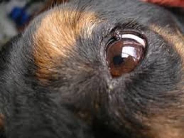
These are common in older dogs and start as small bumps at the margin of the upper and lower eyelids. Prevalence of MGD in. The condition is commonly bilateral but may have a unilateral presentation3 Staphylococcus and Streptococcus species are the isolates most commonly involved in bacterial blepharitis of adult dogs3 In puppies bacterial blepharitis occurs as part of a juvenile pyoderma in which the entire skin. Sebaceous adenitis and concurrent meibomian gland dysfunction MGD were diagnosed in a two-year-old mongrel dog presenting with hypotrichosis exfoliative dermatitis and blepharitis. Sometimes little bumps overgrowths of blocked oil from the Meibomian glands - form into tumors. Cytology and culture of the purulent discharge from one of the meibomian glands yielded Streptococcus species.
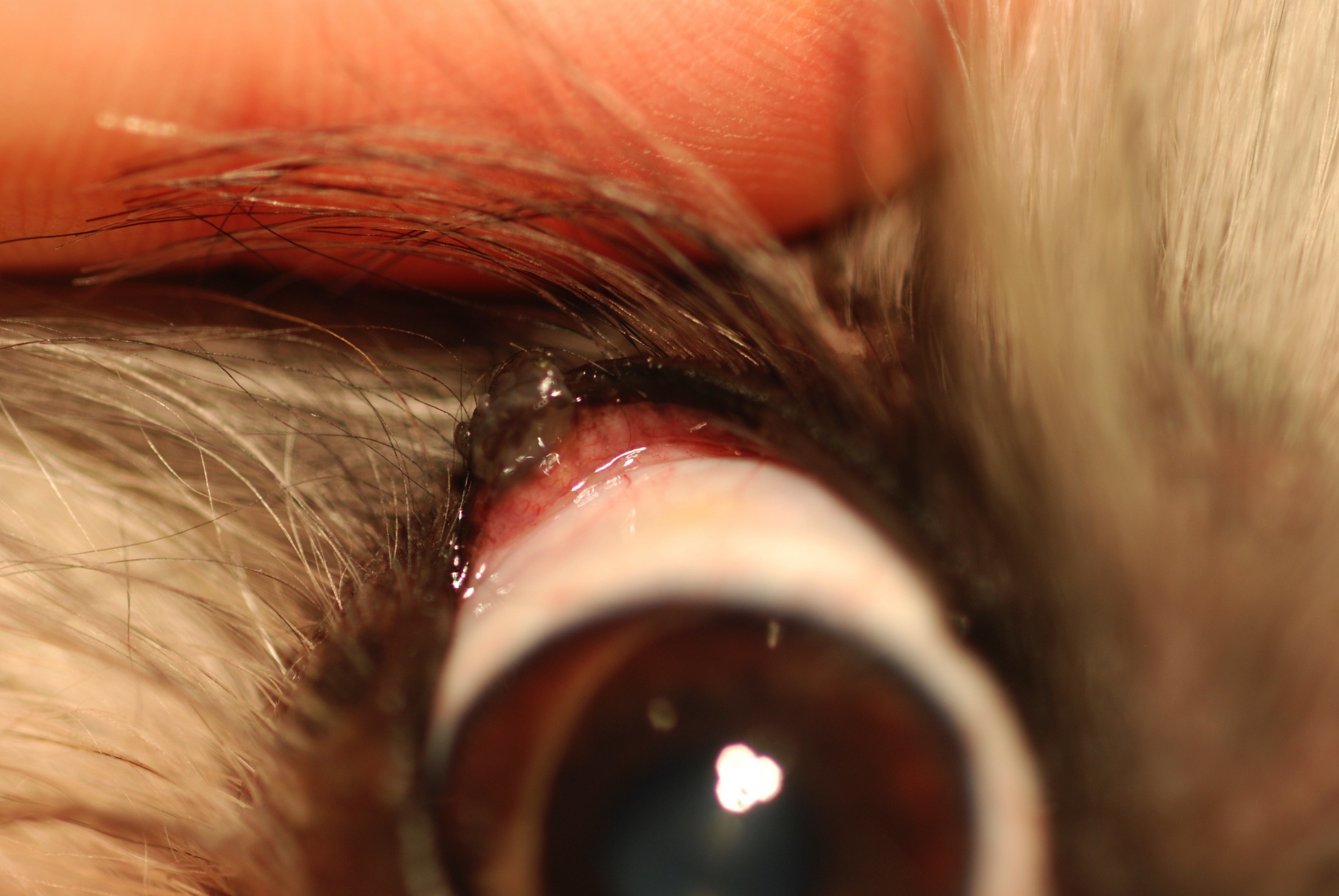
Meibomian Gland Adenomas MGA are benign age related eyelid tumors which result from the accumulation of glandular material.
The eyelids of dogs cats and people contain Meibomian glands. Much of what is known today about the Meibomian gland and MGD was learnt from these important animal models. Health Navigator says these are usually caused by blocked oil glands and they are not due to. Meibomian gland tumors are tiny slow-growing tumors that form in the meibomian glands of the eyelids. These glands become dysfunctional if blocked leading to swelling within or on the margins of the eyelid. Meibomian glands secrete oily substances that help keep the tear film healthy.
 Source: southeasternvet.com.au
A combination of oral antimicrobials a tapering dose of steroids and. These are common in older dogs and start as small bumps at the margin of the upper and lower eyelids. In particular canine and rabbit models have been essential for studying the physiopathology and progression of DED and the mouse model which includes different knockout strains. The tear film protects the eye from particles dirt dust and it also helps to keep the eye moist. When the opening of the meibomian gland duct gets clogged the oil builds up in the gland and causes inflammation.
 Source: urbananimalveterinary.com
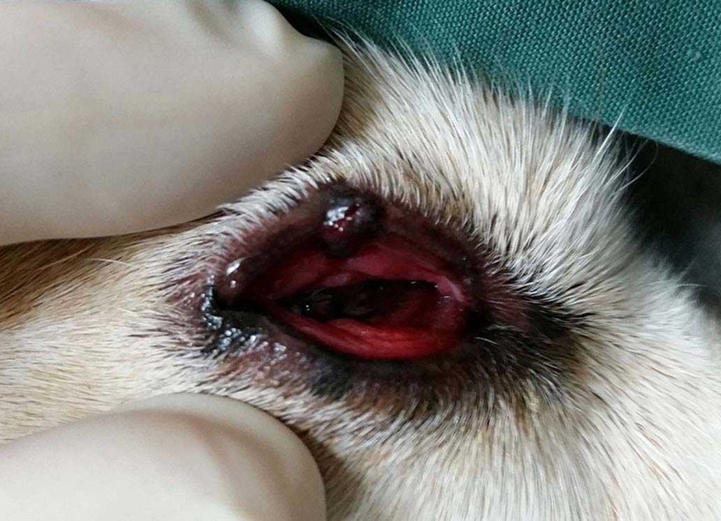
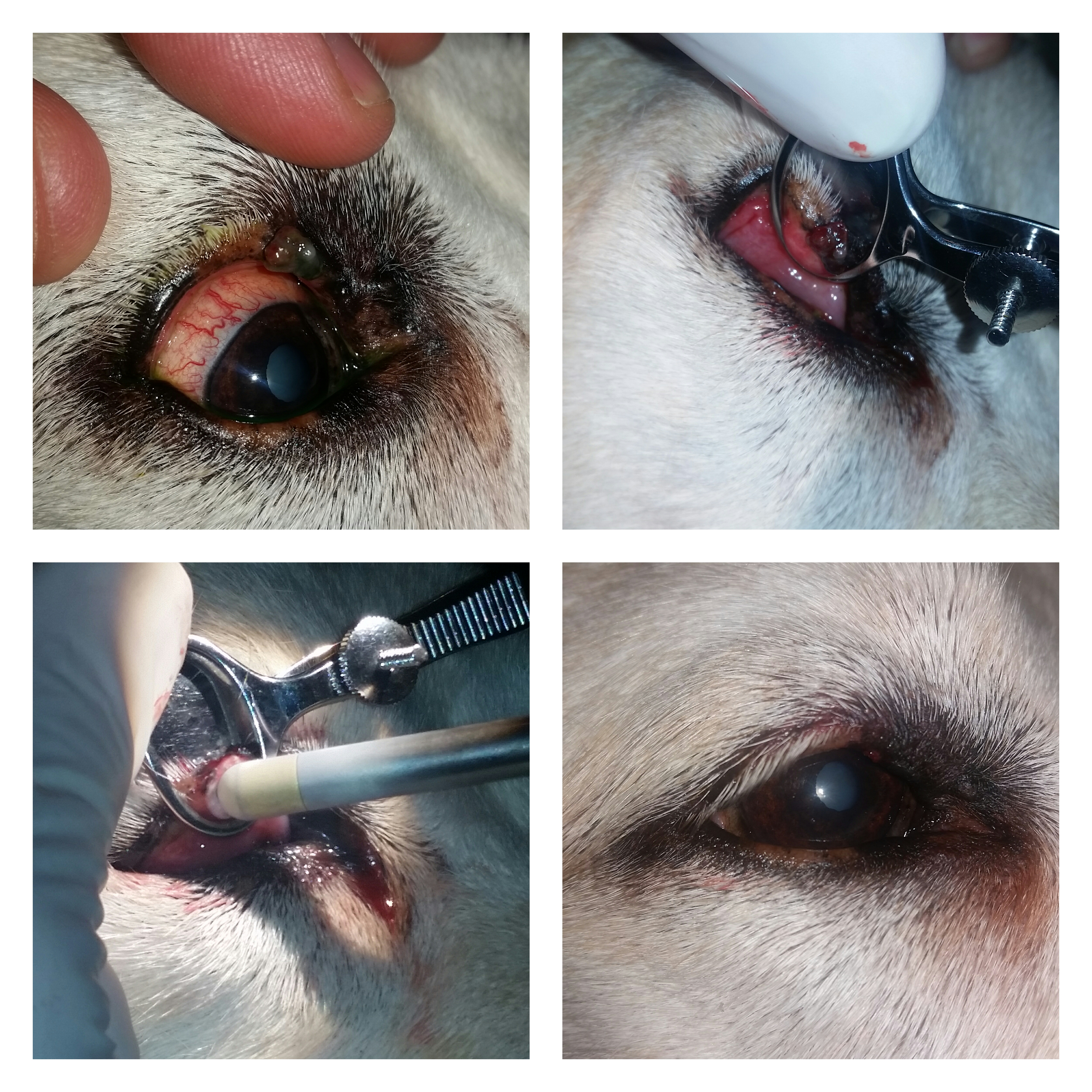
Canine meibomian gland tumors can become irritated painful and even infected or they can cause corneal ulcers or conjunctivitis. Cytology and culture of the purulent discharge from one of the meibomian glands yielded Streptococcus species. Dozens of these glands are in each eyelid. If they become large enough MGAs can cause irritation to the cornea and conjunctiva and may reduce the normal ability to blink. These glands become dysfunctional if blocked leading to swelling within or on the margins of the eyelid.
 Source: acvo.org
Meibomian adenoma and epithelioma represent 10 of tumor submissions to COPLOW and are the most frequent eyelid tumors in dogs of middle to advanced age comprising 44 of canine eyelid tumors Peiffer and Simmons 2002A survey of 202 canine eye lid tumors found that histopathologically 44 were sebaceous gland and benign eyelid tumors 733. Meibomian glands secrete oily substances that help keep the tear film healthy. Health Navigator says these are usually caused by blocked oil glands and they are not due to. What Are Meibomian Glands in Dogs. Theyre not underneath the eyelid.
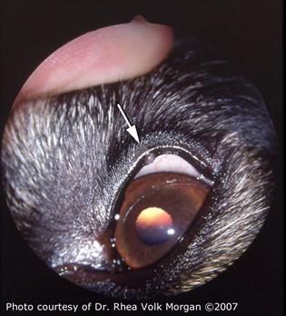
Much of what is known today about the Meibomian gland and MGD was learnt from these important animal models. What Are Meibomian Glands in Dogs. Much of what is known today about the Meibomian gland and MGD was learnt from these important animal models. Two-year-old castrated male mixed breed dog with bacterial blepharitis Streptococcus species. Health Navigator says these are usually caused by blocked oil glands and they are not due to.
 Source: acvo.org
Theyre not underneath the eyelid. Meibomian gland dysfunction MGD is one of the possible conditions underlying ocular surface disorders OSD. These glands become dysfunctional if blocked leading to swelling within or on the margins of the eyelid. Both dog and cat eyes may be affected by these but they are more common in dogs. Two-year-old castrated male mixed breed dog with bacterial blepharitis Streptococcus species.
 Source: mspca.org
Sebum prevents the evaporation of the dogs natural tear film. While these are technically benign in. Both dog and cat eyes may be affected by these but they are more common in dogs. Changes of meibomian glands detected by NIM in dogs12 Here we present a case of localized meibomian gland drop-out as revealed by NIM in the center of the right upper eye-lid in a Cairn terrier with pigmentary glaucoma. In dogs Meibomian gland tumors usually grow slowly.
 Source: eyespecialistsforanimals.com
In older dogs it is quite common for one gland to get a little overexcited and form a growth. Note diffuse ulceration of both eyelids with nodule formation crusting and discharge. Meibomian adenoma and epithelioma represent 10 of tumor submissions to COPLOW and are the most frequent eyelid tumors in dogs of middle to advanced age comprising 44 of canine eyelid tumors Peiffer and Simmons 2002A survey of 202 canine eye lid tumors found that histopathologically 44 were sebaceous gland and benign eyelid tumors 733. What are meibomian gland tumours. Diagnosis of sebaceous adenitis was based on history clinical signs the histological demonstration of multifocal lymphohistiocytic and neutrophilic inflammation targeting the.
 Source: pinterest.com
The eyelids of dogs cats and people contain Meibomian glands. A combination of oral antimicrobials a tapering dose of steroids and. Meibomian gland tumors are tiny slow-growing tumors that form in the meibomian glands of the eyelids. Changes of meibomian glands detected by NIM in dogs12 Here we present a case of localized meibomian gland drop-out as revealed by NIM in the center of the right upper eye-lid in a Cairn terrier with pigmentary glaucoma. It goes without saying that these videos are intended for veterinary prof.
 Source: onlinelibrary.wiley.com
Dozens of these glands are in each eyelid. For such animal models have shown to be a vital tool. It goes without saying that these videos are intended for veterinary prof. Meibomian glands produce sebum oil which helps keep the surface of the eye lubricated. Canine meibomian gland tumors can become irritated painful and even infected or they can cause corneal ulcers or conjunctivitis.
 Source: animalcareinfo.com
While these are technically benign in. It goes without saying that these videos are intended for veterinary prof. Meibomian gland tumors in dogs are usually benign non-cancerous so they dont typically spread or move to other areas of the body. Meibomian gland tumors are tiny slow-growing tumors that form in the meibomian glands of the eyelids. A combination of oral antimicrobials a tapering dose of steroids and.
 Source: msdvetmanual.com
Both dog and cat eyes may be affected by these but they are more common in dogs. Sometimes little bumps overgrowths of blocked oil from the Meibomian glands - form into tumors. In older dogs it is quite common for one gland to get a little overexcited and form a growth. Sebum prevents the evaporation of the dogs natural tear film. Meibomian gland dysfunction MGD is one of the possible conditions underlying ocular surface disorders OSD.
 Source: msdvetmanual.com
Dozens of these glands are in each eyelid. If you notice a growth on your dogs eyelid it could be whats known as a meibomian gland cyst or chalazion. Canine meibomian gland tumors can become irritated painful and even infected or they can cause corneal ulcers or conjunctivitis. Changes of meibomian glands detected by NIM in dogs12 Here we present a case of localized meibomian gland drop-out as revealed by NIM in the center of the right upper eye-lid in a Cairn terrier with pigmentary glaucoma. In dogs Meibomian gland tumors usually grow slowly.
If you find this site good, please support us by sharing this posts to your favorite social media accounts like Facebook, Instagram and so on or you can also bookmark this blog page with the title meibomian gland dog by using Ctrl + D for devices a laptop with a Windows operating system or Command + D for laptops with an Apple operating system. If you use a smartphone, you can also use the drawer menu of the browser you are using. Whether it’s a Windows, Mac, iOS or Android operating system, you will still be able to bookmark this website.


