Thick intestinal wall in dogs
Thick Intestinal Wall In Dogs. Note the marked difference in overall wall thickness between the small SI. Ultrasound of the gastrointestinal GI tract has become an increasingly popular and useful diagnostic procedure for evaluation of gastric and intestinal disease. The normal sonographic thickness of the individual layers ie mucosa submucosa muscularis and subserosa-serosa of the intestinal wall was evaluated in 20 clinically healthy cats. The duodenum is visible in the near field SI.
 Ultrasound Cross Scan Of Stomach Wall In A 12 Years Old Dog A Focal Download Scientific Diagram From researchgate.net
Leiomyosarcomas are considered the second most common canine intestinal tumor and the most common intestinal sarcoma in dogs. A The major duodenal papilla in the duodenum of the dog is the site of entry of the common bile duct and major pancreatic ductThe minor duodenal papilla is seen in some but not all dogs distal to the major papilla and approximately 100 degrees clockwise from itB Normal descending. She has had no vomiting diarrhea or weight loss. If the ulcer extends through the stomach or intestinal wall food and digestive fluids can escape into the abdomen. Approach to the thickened bowel wall. The intestine is most commonly sutured using a full thickness simple continuous or simple interrupted pattern and swaged on suture material.
2 - 3 mm 6.
Other forms of gastritis that cause a thickening include eosinophilic gastritis which causes white blood cells to gather on the walls of the dogs stomach. A positive association between intestinal wall thickness in dogs and either the histological diagnosis or the response to treatment was not found. The intestine is most commonly sutured using a full thickness simple continuous or simple interrupted pattern and swaged on suture material. The duodenum is visible in the near field SI. A vet will rely on blood work imaging and biopsies to determine the cause of a thickened stomach wall. It is not necessary to use inverting suture patterns nor to do a two-layer closure.
 Source: researchgate.net
Increased thickness of the intestinal wall is a hallmark for the detection of diseases ranging from inflammatory bowel disease to neoplasia. One wall highlighted intestine. Ultrasonographic intestinal wall measurements do not appear to be able to establish a diagnosis of intestinal inflammation and may result in a false negative diagnosis in cases of IBD. The duodenum is visible in the near field SI. A vet will rely on blood work imaging and biopsies to determine the cause of a thickened stomach wall.
 Source: vetgrad.com
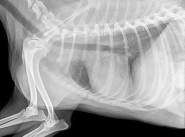
Conclusions and Clinical Relevance Values for thickness of the wall layers of the duodenum jejunum and colon of dogs reported here may be useful for assessing gastrointestinal tract diseases primarily targeting a specific. She has had no vomiting diarrhea or weight loss. It is not necessary to use inverting suture patterns nor to do a two-layer closure. Other diagnostic procedures include abdominal X-rays and ultrasounds which help to identify changes in the stomach and intestinal walls. Stomach tumors and cancer may also cause a thickening of the stomach wall.
 Source: researchgate.net
Stomach tumors and cancer may also cause a thickening of the stomach wall. A thickened stomach lining in dogs is usually caused by a bout of gastritis or pyloric obstruction also referred to as stenosis. Normal bowel wall is 30 to 50 mm thick in adults6 Our study done on a small number of normal dogs showed a range of 30 to 50 for the stomach and 20. Segment of Gastrointestinal Tract Dog Wall Thickness Cat Wall Thickness. The walls of the tube are composed of glands nerves and muscles.
Source: researchgate.net
An ultra sound this morning showed no masses normal kidneys spleen gall bladder liver lymph nodes. Ultrasonographic intestinal wall measurements do not appear to be able to establish a diagnosis of intestinal inflammation and may result in a false negative diagnosis in cases of IBD. The walls of the tube are composed of glands nerves and muscles. Pyloric Obstruction or Stenosis Pyloric obstruction also known as stenosis occurs when the layers of the dogs stomach lining become thick and inflamed. 15 - 2 mm 8-10.
 Source: vetgrad.com
2 - 25 mm 67. The mean thickness of the wall was 220 222 300 and 204 mm for duodenum jejunum ileum fold and ileum between folds respectively. Figure 57-2 Videoendoscopic appearance of the normal upper small intestine. Normal bowel wall is 30 to 50 mm thick in adults6 Our study done on a small number of normal dogs showed a range of 30 to 50 for the stomach and 20. The duodenum is visible in the near field SI.
 Source: researchgate.net
26 Leiomyosarcoma causes large eccentrically located single or multiple hypoechoic or anechoic areas of wall thickening. Your biopsy site should be prepared as described in the previous section Place a full-thickness 3-0 or 4-0 stay suture through the antimesenteric wall at the site you would like to sample. 3 - 5 mm 2. Up to 5 mm 5. A positive association between intestinal wall thickness in dogs and either the histological diagnosis or the response to treatment was not found.
 Source: semanticscholar.org
25 - 32 mm 467. However the outlook is poor for those with ulcers associated with renal or liver failure and for animals with cancers such as stomach carcinoma and gastrinoma. It is not necessary to use inverting suture patterns nor to do a two-layer closure. Increased thickness of the intestinal wall is a hallmark for the detection of diseases ranging from inflammatory bowel disease to neoplasia. Your biopsy site should be prepared as described in the previous section Place a full-thickness 3-0 or 4-0 stay suture through the antimesenteric wall at the site you would like to sample.
 Source: todaysveterinarypractice.com
Ultrasonographic intestinal wall measurements do not appear to be able to establish a diagnosis of intestinal inflammation and may result in a false negative diagnosis in cases of IBD. Other diagnostic procedures include abdominal X-rays and ultrasounds which help to identify changes in the stomach and intestinal walls. Your biopsy site should be prepared as described in the previous section Place a full-thickness 3-0 or 4-0 stay suture through the antimesenteric wall at the site you would like to sample. One cadaveric study suggested avoiding the use of conventional cutting needles Mitsou et al 2018. Ultrasound of the gastrointestinal GI tract has become an increasingly popular and useful diagnostic procedure for evaluation of gastric and intestinal disease.
 Source: todaysveterinarypractice.com
2 mm inter-rugal 34 and 4 mm rugal fold thickness 3. Mean SD thickness of the colonic wall for small medium and large dogs was 15 03 mm 14 05 mm and 16 04 mm respectively. Pyloric Obstruction or Stenosis Pyloric obstruction also known as stenosis occurs when the layers of the dogs stomach lining become thick and inflamed. But none of the other symptoms of lymphsarcoma. My dog has thickened intestinal walls low energy and loss of appetite.
 Source: vetgrad.com
A vet will rely on blood work imaging and biopsies to determine the cause of a thickened stomach wall. One wall highlighted intestine. The intestine is most commonly sutured using a full thickness simple continuous or simple interrupted pattern and swaged on suture material. 2 mm inter-rugal 34 and 4 mm rugal fold thickness 3. Conclusions and Clinical Relevance Values for thickness of the wall layers of the duodenum jejunum and colon of dogs reported here may be useful for assessing gastrointestinal tract diseases primarily targeting a specific.
 Source: todaysveterinarypractice.com
26 Leiomyosarcoma causes large eccentrically located single or multiple hypoechoic or anechoic areas of wall thickening. 2 - 4 mm 6. Other forms of gastritis that cause a thickening include eosinophilic gastritis which causes white blood cells to gather on the walls of the dogs stomach. Increased thickness of the intestinal wall is a hallmark for the detection of diseases ranging from inflammatory bowel disease to neoplasia. 26 Leiomyosarcoma causes large eccentrically located single or multiple hypoechoic or anechoic areas of wall thickening.
 Source: vetgrad.com
Ultrasonographic intestinal wall measurements do not appear to be able to establish a diagnosis of intestinal inflammation and may result in a false negative diagnosis in cases of IBD. The walls of the tube are composed of glands nerves and muscles. One wall highlighted intestine. Conclusions and Clinical Relevance Values for thickness of the wall layers of the duodenum jejunum and colon of dogs reported here may be useful for assessing gastrointestinal tract diseases primarily targeting a specific. Segment of Gastrointestinal Tract Dog Wall Thickness Cat Wall Thickness.
If you find this site convienient, please support us by sharing this posts to your preference social media accounts like Facebook, Instagram and so on or you can also bookmark this blog page with the title thick intestinal wall in dogs by using Ctrl + D for devices a laptop with a Windows operating system or Command + D for laptops with an Apple operating system. If you use a smartphone, you can also use the drawer menu of the browser you are using. Whether it’s a Windows, Mac, iOS or Android operating system, you will still be able to bookmark this website.